How Do Doctors See All Parts in a Pregnancy Scan? 🤰🩺
🌼 First things first: Ultrasound is safe—and thorough
Pregnancy ultrasound uses high-frequency sound waves—not radiation—to create moving images of your baby. With the right angles and image settings, doctors can see the head and brain structures, face, heart, spine, abdomen, kidneys, stomach, bladder, limbs, placenta, and umbilical cord, plus amniotic fluid and overall growth.
Good to know: Sometimes getting all these views in one go takes patience—baby’s position matters!
Watch our Reel on :How Do Doctors See All Parts in a Pregnancy Scan?
🧭 What exactly are doctors looking at?
During a routine scan, the sonologist/fetal medicine specialist follows a structured checklist so nothing important is missed.
Head & Brain: skull shape, midline structures, ventricles, cerebellum, posterior fossa
Face: profile (including nasal bone), orbits (eye lenses), lips and nose
Heart (Fetal Echo Views): four-chamber view, outflow tracts, three-vessel view, valve flows (as needed)
Spine: neck to sacrum in both longitudinal and transverse planes
Chest & Lungs: symmetry, heart position
Abdomen: stomach bubble, liver, bowel, abdominal wall integrity
Kidneys & Bladder: both kidneys visible? bladder filling?
Limbs: arms, forearms, hands; thighs, legs, feet; basic movements
Placenta & Cord: location (anterior/posterior/fundal/low-lying), cord insertion, Doppler if indicated
Amniotic Fluid: pockets and overall index
Growth: measurements like BPD, HC, AC, FL, and estimated fetal weight (EFW)
This protocol helps ensure the scan is consistent and comprehensive—even if baby is playful, shy, or super sleepy that day!
📷 How can sound waves “see” inside?
The ultrasound probe sends sound waves that bounce off tissues at different speeds. The machine converts echoes into a grayscale image.
- Bright (echogenic): bone (like skull or spine)
- Medium gray: soft tissues (organs, muscles)
- Dark/black: fluid (amniotic fluid, bladder, stomach contents)
By tilting, rotating, and sliding the probe, the doctor captures multiple planes (longitudinal, transverse, coronal) to build a full picture of every part.
🧒 Why do doctors ask me to walk, drink water, eat something sweet, or cough?
Great question—and totally normal to wonder!
- Walk around / change position: helps baby turn so hidden parts (like heart outflow tracts or facial profile) become visible.
- Drink water (early pregnancy): a comfortably full bladder lifts the uterus and improves early views.
- Eat something sweet (later pregnancy): gentle fetal activity can improve views when baby is too sleepy or stuck in one position.
- Cough / shift / left-right tilt: tiny movements change acoustic windows, allowing clearer images of tricky areas.
👉 These are techniques to improve visibility, not signs that something is wrong. Please don’t worry if your doctor asks you to try them—they’re part of getting a perfect, complete study.
🗓️ Which scan checks what (and when)?
1) Early/Dating Scan (around 6–10 weeks)
- Confirms pregnancy location (inside uterus), single/twin, viability (heartbeat), and dates.
2) NT Scan (11–13+6 weeks)
- Nuchal translucency, nasal bone, early anatomy overview, some blood-flow markers; combined with maternal bloods if doing first-trimester screening.
3) Anomaly Scan (18–22 weeks)
- The big, detailed survey of every organ system. This is where doctors methodically go through head-to-toe anatomy.
4) Growth/Well-being Scans (third trimester as advised)
- Tracks growth, fluid, placenta, and often includes BPP/Dopplers if needed.
🧩 Why sometimes a “repeat view” or “follow-up scan”?
Even with expertise and advanced machines, certain views can be temporarily difficult because of:
- Fetal position (back facing the probe, chin tucked, legs crossed)
- Low-lying placenta or maternal abdominal wall thickness
- Very active or very sleepy baby
- Gestational age (some structures are best seen at specific weeks)
A short repeat attempt after a break—or a follow-up appointment—is common and benefits accuracy. The goal is a complete, confident report.
❤️ How doctors examine the fetal heart
The heart deserves special attention. Doctors assess:
- Four-chamber view (size, symmetry, septum)
- Outflow tracts (aorta, pulmonary artery)
- Three-vessel/three-vessel-trachea view
- Rhythm and valve flows (if indicated)
If any view isn’t crystal clear, they may try color Doppler, ask you to change sides, or re-check after a brief walk. Again—this is about precision, not panic.
👶 Limbs, fingers, toes—how do they check?
Limb bones (humerus, radius/ulna, femur, tibia/fibula) are measured and visualized in two planes. Hands and feet are counted/seen; movements reassure good tone and neuromuscular development. Sometimes little fists stay clenched—doctors may wait for a natural open-close moment to confirm digits.
🧪 What if the report mentions “limited view” of a part?
That line usually means the view wasn’t optimal due to position or shadowing. The standard response is to repeat the specific view, often the same day after a short break, or in a few days. It does not automatically mean a problem—just that the team wants better pictures before signing off.
🧘 Preparation tips to help your scan go smoother
- Arrive on time and relaxed—stress can make the experience feel longer.
- Follow bladder instructions (early scans: comfortably full; later scans: usually normal bladder).
- Carry past reports and doctor’s notes.
- Light snack (as advised) can encourage gentle fetal movement in later scans.
- Wear comfy clothing for easy abdominal access.
🧑⚕️ Who is doing your scan matters
-
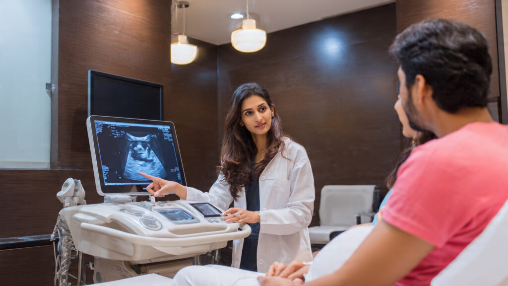
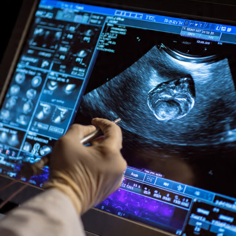
At centers like Nisarga Diagnostics (Sanjaynagar, Bengaluru), scans are performed by FMF-certified Fetal Medicine Specialists using high-resolution machines. The combination of expertise + protocol + patience is what lets doctors truly “see all parts” and counsel you with confidence and compassion.
Common worries—answered
“Doctor asked me to walk/eat sweets—should I worry?”
No—these are standard maneuvers to get perfect views.“Why did my scan take longer than my friend’s?”
Every baby is unique! Position, movements, and individual anatomy affect scan time. Longer ≠ worse—often it means your team is being thorough.“Is ultrasound harmful?”
Diagnostic obstetric ultrasound is considered safe when used appropriately by trained professionals.
📌 Key takeaways
-
- Ultrasound is a safe, structured, full-body check of your baby and pregnancy.
- Doctors use multiple angles and planes to assess each organ.
- Requests to walk/drink water/eat something sweet/cough are simple tricks to improve views.
- Repeat views ensure accuracy, not alarm.
- Choose an experienced, well-equipped center for the best experience.
📲 Book your scan with confidence
-
-
Nisarga Diagnostics, Sanjaynagar, Bengaluru
+91 88677 57594 | +91 91025 97025
Open all days — Sundays & festivals included
FMF-certified Fetal Medicine Specialists • Advanced Ultrasound
-

Founder of Nisarga Diagnostics, is a renowned fetal ultrasound specialist with over 20 years of expertise. An alumnus of Government Medical Colleges in Mysore and Bangalore, he also holds a D.N.B. from the National Board of Examinations, Delhi, and certification from the Fetal Medicine Foundation, London. A former Professor and HOD, he has trained countless PG students and junior radiologists, shaping the future of fetal imaging in India.
