

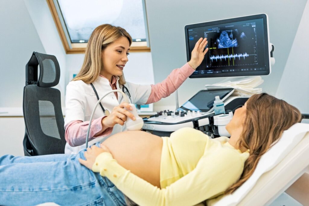
Fetal Echo Scan: Complete Guide to Your Baby's Heart Scan During Pregnancy
Pregnancy is a remarkable journey, and every parent hopes for a healthy baby. Among the many prenatal scans available, one specialized test focuses exclusively on the baby’s heart—the Fetal Echocardiography (Fetal Echo) Scan.
A fetal echo provides a detailed evaluation of the baby’s heart while still in the womb. It helps doctors identify congenital heart defects, heart rhythm abnormalities, and other cardiac conditions before birth, allowing for timely monitoring and appropriate care.
At Nisarga Diagnostics, our advanced fetal echocardiography service provides accurate assessment of your baby’s heart using high-resolution ultrasound technology, helping expectant parents receive early answers and peace of mind.
Watch our video on : What is a Fetal Echo Scan?
What is a Fetal Echo Scan?
A Fetal Echo Scan, also called Fetal Echocardiography, is a specialized ultrasound examination that evaluates the baby’s heart in great detail.
Unlike a routine pregnancy scan, which checks the baby’s overall growth and development, a fetal echo focuses specifically on:
- Heart chambers
- Heart valves
- Blood flow
- Major blood vessels
- Heart rhythm
- Electrical conduction
- Overall heart function
This examination is completely safe for both mother and baby because it uses sound waves and does not involve radiation.
Why is a Fetal Echo Scan Done?
-
A fetal echo helps detect heart problems before birth.
It is recommended to:
- Detect congenital heart defects
- Evaluate abnormal heart rhythms
- Assess the baby’s heart structure
- Monitor high-risk pregnancies
- Plan treatment before and after delivery if required
Early diagnosis helps healthcare providers prepare the best possible care for the baby.
Who Needs a Fetal Echo Scan?
Your doctor may recommend a fetal echo if:
Maternal Risk Factors
- Diabetes before pregnancy
- Autoimmune diseases such as lupus
- Positive Anti-Ro (SSA) antibodies
- Positive Anti-La (SSB) antibodies
- Certain medications during pregnancy
- Family history of congenital heart disease
Baby-Related Risk Factors
- Abnormal anomaly scan
- Irregular fetal heartbeat
- Slow fetal heart rate
- Suspected congenital heart disease
- Increased nuchal translucency (NT)
Even if there are no risk factors, a fetal echo may be recommended if your doctor wants a more detailed assessment of the baby’s heart.
When is a Fetal Echo Scan Performed?
The ideal time is:
18 to 24 weeks of pregnancy
In some high-risk pregnancies, it may also be performed earlier or repeated later depending on the baby’s condition.
What Does a Fetal Echo Examine?
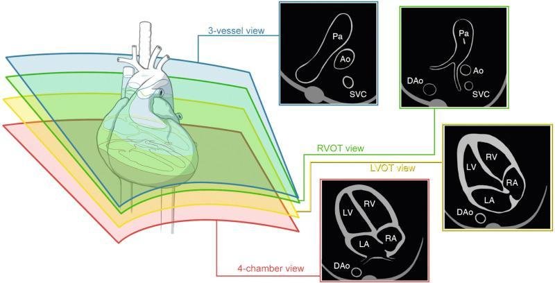
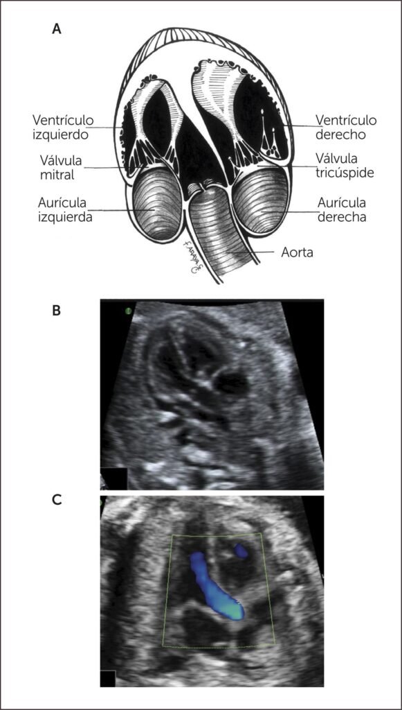
The scan carefully evaluates:
Heart Chambers
- Right atrium
- Left atrium
- Right ventricle
- Left ventricle
Heart Valves
Doctors examine whether the valves open and close properly.
Septum
The scan checks for holes between the heart chambers, such as:
- Atrial Septal Defect (ASD)
- Ventricular Septal Defect (VSD)
Major Blood Vessels
The scan evaluates:
- Aorta
- Pulmonary artery
- Great vessel connections
Heart Rhythm
Doctors ensure the heartbeat is regular and the electrical signals are functioning normally.
Blood Flow
Using Color Doppler imaging, specialists assess how blood moves through the baby’s heart.
What Conditions Can a Fetal Echo Detect?
A fetal echo can identify:
- Congenital heart defects
- Heart valve abnormalities
- Septal defects (holes in the heart)
- Tetralogy of Fallot
- Transposition of the Great Arteries
- Coarctation of the Aorta
- Hypoplastic Left Heart Syndrome
- Fetal heart block
- Cardiac tumors (rare)
- Abnormal heart rhythm (Arrhythmias)
Not every abnormality requires surgery after birth. Some conditions are mild, while others may need specialized treatment.
How is the Scan Performed?
The procedure is simple and painless.
- You lie comfortably on the examination bed.
- Ultrasound gel is applied to your abdomen.
- A specialized ultrasound probe is moved over the abdomen.
- Multiple views of the baby’s heart are obtained.
- The scan usually takes 30–60 minutes, depending on the baby’s position.
Is the Scan Safe?
Yes.
A fetal echo:
- Uses ultrasound only
- Has no radiation
- Is non-invasive
- Is painless
- Is considered safe for both mother and baby
How Should You Prepare?
Usually, no special preparation is required.
You may be advised to:
- Wear comfortable clothing
- Carry previous pregnancy scan reports
- Bring referral documents if available
- Arrive a little early for registration
What Happens if a Heart Problem is Found?
Finding a heart problem early allows doctors to:
- Monitor the pregnancy more closely
- Refer you to a fetal medicine specialist
- Plan delivery at an appropriate hospital
- Arrange pediatric cardiology consultation
- Prepare treatment immediately after birth if needed
Early diagnosis often leads to better planning and improved outcomes.
Frequently Asked Questions (FAQs)
Is a fetal echo different from a routine pregnancy scan?
Yes. A routine scan evaluates the baby’s overall growth and anatomy, while a fetal echo focuses specifically on the heart’s structure and function.
Does every pregnant woman need a fetal echo?
Not necessarily. It is usually recommended when there are risk factors or concerns about the baby’s heart. Your obstetrician will advise if it is needed.
Can a fetal echo detect all heart problems?
A fetal echo can detect many major congenital heart defects and rhythm abnormalities. However, some minor conditions may only become apparent after birth.
How long does a fetal echo take?
The scan typically takes 30 to 60 minutes, depending on the baby’s position and the complexity of the examination.
Is a fetal echo painful?
No. It is a painless, non-invasive ultrasound procedure that is safe for both mother and baby.
Conclusion
A Fetal Echo Scan is one of the most important specialized investigations when there is concern about a baby’s heart during pregnancy. It provides detailed information about the heart’s structure, rhythm, and function, allowing doctors to detect congenital heart conditions early and plan appropriate care.
If your obstetrician has recommended a fetal echo, timely evaluation can provide valuable information and reassurance.
At Nisarga Diagnostics, we are dedicated to delivering advanced fetal heart imaging with precision, expertise, and compassionate care to support healthy pregnancies.

Founder of Nisarga Diagnostics, is a renowned fetal ultrasound specialist with over 20 years of expertise. An alumnus of Government Medical Colleges in Mysore and Bangalore, he also holds a D.N.B. from the National Board of Examinations, Delhi, and certification from the Fetal Medicine Foundation, London. A former Professor and HOD, he has trained countless PG students and junior radiologists, shaping the future of fetal imaging in India.