
NT Scan During Pregnancy: Timing, Purpose & Results
Pregnancy involves several important scans that help monitor your baby’s growth and development. One of the most crucial first-trimester scans is the NT Scan During Pregnancy. This scan helps doctors assess your baby’s development and estimate the risk of certain chromosomal abnormalities and pregnancy-related complications.
In this article, we explain the purpose of an NT Scan the ideal time to undergo the scan, what it detects, and what the results mean.
Watch our video on : NT Scan During Pregnancy: Timing, Purpose & Results
What Is an NT Scan During Pregnancy?
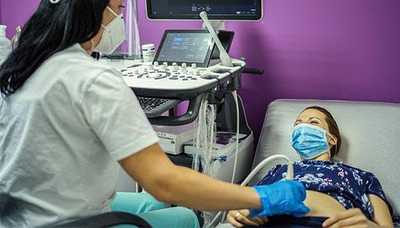
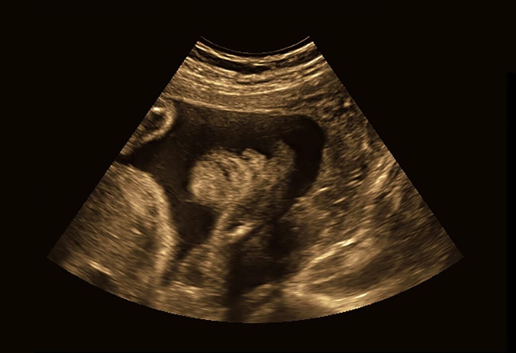
An Nuchal Translucency Scan is a specialized ultrasound performed during the first trimester. It measures the fluid-filled space at the back of the baby’s neck, known as the nuchal translucency.
The measurements obtained during the scan are combined with maternal information and specialized software calculations to estimate the risk of certain chromosomal conditions.
Unlike routine pregnancy scans, an NT Scan must be performed within a specific timeframe for accurate results.
When Should an NT Scan During Pregnancy Be Done?
-
The ideal time for an NT Scan is between:
11 weeks 2 days and 14 weeks 1 day
This is an internationally accepted guideline followed worldwide. The scan cannot be accurately interpreted before or after this period because the specialized risk-calculation software is validated only within this gestational age window.
Missing this timeframe may limit the usefulness of the test.
Why Is an NT Scan During Pregnancy Important?
An NT Scan provides valuable information about both the baby and the pregnancy.
The scan helps:
- Confirm gestational age
- Assess fetal growth
- Detect major structural abnormalities
- Estimate the risk of chromosomal abnormalities
- Assess the risk of certain pregnancy complications
Early identification of potential concerns allows doctors to recommend appropriate monitoring and treatment.
What Does the Radiologist Evaluate During an NT Scan During Pregnancy?
1. Baby’s Growth and Gestational Age
The baby’s measurements are assessed to confirm the number of weeks of pregnancy and ensure normal development.
2. Major Structural Abnormalities
An NT Scan During Pregnancy can identify certain major abnormalities involving:
- Skull development
- Brain formation
- Spine development
- Limbs and extremities
Although not every condition can be detected at this stage, significant abnormalities may be identified early.
3. Risk of Chromosomal Abnormalities
One of the main purposes of an NT Scan During Pregnancy is to estimate the risk of chromosomal disorders.
These include:
Trisomy 21 (Down Syndrome)
Down Syndrome occurs when there is an extra copy of chromosome 21.
Trisomy 18
A chromosomal disorder associated with serious developmental abnormalities.
Trisomy 13
A rare but severe chromosomal condition that affects multiple organs.
It is important to understand that an NT Scan During Pregnancy does not diagnose these conditions. Instead, it calculates the likelihood or risk that they may be present.
Can an NT Scan During Pregnancy Predict Future Pregnancy Complications?
Yes. In addition to chromosomal screening, an NT Scan During Pregnancy may help estimate the risk of:
Preeclampsia
Preeclampsia is a pregnancy-related condition characterized by high blood pressure and can affect both mother and baby.
Intrauterine Growth Restriction (IUGR)
IUGR refers to poor fetal growth during pregnancy.
Reduced Fetal Weight Gain in Later Pregnancy
The scan may help identify pregnancies that require closer monitoring during the third trimester.
How Should You Prepare for an NT Scan During Pregnancy?
Before attending your scan appointment, carry:
- Doctor’s prescription
- Previous pregnancy scan reports
- Dating Scan report
- Pregnancy records
- Original identification document
Having previous reports available helps the radiologist compare findings and provide a more accurate assessment.
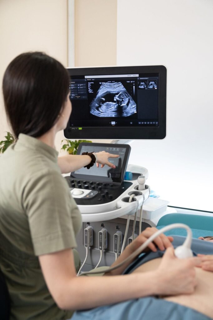
What Information Is Included in an NT Scan Report?
An NT Scan During report usually includes:
- Gestational age
- Fetal measurements
- Assessment of major structural abnormalities
- Risk estimation for chromosomal abnormalities
- Risk assessment for preeclampsia
- Risk assessment for fetal growth restriction
The report typically classifies risk as either low risk or high risk.
Low-Risk Result
A low-risk result suggests a lower probability of chromosomal abnormalities and is generally reassuring.
High-Risk Result
A high-risk result does not mean that the baby definitely has a chromosomal abnormality. It only indicates an increased probability and may require further evaluation.
Your doctor may recommend additional screening or diagnostic tests if necessary.
What Happens After an NT Scan During Pregnancy?
After receiving the report, you should consult your obstetrician or gynecologist.
If the findings are normal, routine pregnancy care will continue as planned.
The next important scan is usually the Anomaly Scan, which is performed between 18 and 20 weeks of pregnancy to evaluate the baby’s organs and anatomy in greater detail.
If the NT Scan indicates an increased risk of chromosomal abnormalities, preeclampsia, or fetal growth restriction, your doctor may suggest additional monitoring and preventive treatment.
Conclusion
An NT Scan is one of the most important first-trimester scans. It helps evaluate your baby’s growth, detect major abnormalities, assess chromosomal risk, and identify pregnancies that may require closer monitoring.
Because the scan can only be performed between 11 weeks 2 days and 14 weeks 1 day, it is important to schedule it within the recommended timeframe.
Early screening helps ensure timely medical guidance and contributes to a healthier pregnancy journey.

Founder of Nisarga Diagnostics, is a renowned fetal ultrasound specialist with over 20 years of expertise. An alumnus of Government Medical Colleges in Mysore and Bangalore, he also holds a D.N.B. from the National Board of Examinations, Delhi, and certification from the Fetal Medicine Foundation, London. A former Professor and HOD, he has trained countless PG students and junior radiologists, shaping the future of fetal imaging in India.